


MBBS, MS - General Surgery, M.Ch – Urology
A 70-Year-Old's Battle Against Advanced Kidney Cancer - Paras Health, Panchkula
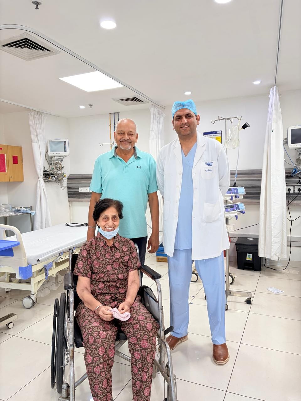
In a remarkable example of advanced cancer surgery and teamwork, Dr Dushiant Sharma and Dr Karan Midha successfully treated a 70-year-old woman suffering from an aggressive right kidney tumor that had extended into one of the body’s largest veins, the inferior vena cava (IVC). Such...
In a remarkable example of advanced cancer surgery and teamwork, Dr Dushiant Sharma and Dr Karan Midha successfully treated a 70-year-old woman suffering from an aggressive right kidney tumor that had extended into one of the body’s largest veins, the inferior vena cava (IVC). Such cases are considered among the most technically challenging operations in urology and cancer surgery.
The patient had been experiencing weakness, reduced appetite, and abdominal discomfort before medical investigations revealed a large tumor in the right kidney. Pre-operative scans showed that the cancer had spread into the IVC, the major vein carrying blood back to the heart. According to imaging, the tumor thrombus had reached a high level near the liver, making the surgery extremely high risk.
A multidisciplinary surgical team planned the operation carefully, anticipating the need for advanced vascular and liver-related surgical techniques. During surgery, doctors used Transesophageal Echocardiography (TEE), a specialized ultrasound performed through the food pipe, to continuously monitor the upper extent of the clot inside the vein. This technology is critical in such operations because it helps surgeons assess the exact location of the tumor thrombus and reduces the risk of life-threatening complications such as clot migration to the heart or lungs.
To safely expose the large abdominal vein, Dr Karan Midha performed complete liver mobilization, a highly specialized step usually reserved for complex cancer surgeries involving major blood vessels. After careful dissection and direct Doppler ultrasound assessment over the IVC , surgeons discovered that the clot was actually located lower than initially suggested on scans. Instead of a higher Level III thrombus, it was found to be a Level I thrombus during surgery.
This important intraoperative finding allowed the surgeons to modify the surgical plan and avoid even more extensive procedures that may have required cardiac bypass support. The team then successfully performed a right radical nephrectomy, removing the cancerous kidney along with complete removal of the tumor clot from the IVC.
The surgery was completed successfully with restoration of normal blood flow in the vein. The patient remained stable after surgery and was shifted for close postoperative monitoring.
Dr Dushiant mentions that kidney cancers extending into major veins require highly experienced surgical teams, advanced imaging support, and careful coordination between urologists, anesthesiologists, GI surgeons ,CTVS surgeons, and critical care specialists. These operations carry significant risks including major bleeding, pulmonary embolism, and cardiac complications.
Dr Dushiant emphasised that this case highlights how modern surgical technology, real-time imaging, and multidisciplinary expertise can help successfully manage even highly advanced kidney cancers. It also underlines the importance of early diagnosis and timely referral to specialized urology centers for patients with complex renal tumors.